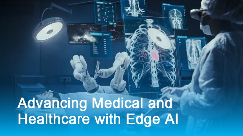
The breakthrough in AI medical image segmentation technology has reached unprecedented scales, with advanced systems now processing 200,000 CT layers daily whilst maintaining 100% DICOM support compatibility. This revolutionary advancement represents a quantum leap in diagnostic imaging capabilities, enabling healthcare professionals to analyse complex medical scans with remarkable speed and precision. The integration of artificial intelligence with medical image segmentation has transformed how radiologists, surgeons, and medical researchers approach patient diagnosis, treatment planning, and clinical research, creating new possibilities for early disease detection and personalised healthcare solutions.
Understanding AI-Powered Medical Image Processing
The magic behind AI medical image segmentation lies in its ability to automatically identify and isolate specific anatomical structures within medical images ??. Unlike traditional manual segmentation methods that could take hours or even days, AI-powered systems can process thousands of CT layers in minutes whilst maintaining clinical-grade accuracy.
What makes this technology particularly impressive is its deep learning architecture, which has been trained on millions of medical images from diverse patient populations. The system can recognise subtle variations in tissue density, organ boundaries, and pathological changes that might be challenging for human observers to detect consistently. This capability becomes especially crucial when dealing with complex cases involving multiple organ systems or rare conditions.
DICOM Compatibility and Healthcare Integration
The 100% DICOM support represents a game-changer for healthcare institutions worldwide ??. DICOM (Digital Imaging and Communications in Medicine) is the global standard for medical imaging data, and seamless compatibility ensures that medical image segmentation systems can integrate directly into existing hospital workflows without requiring costly infrastructure changes.
This compatibility extends beyond simple file format support. The AI system can automatically extract metadata, patient information, and imaging parameters from DICOM files, enabling intelligent processing decisions based on scan protocols, patient demographics, and clinical context. Healthcare professionals can maintain their familiar workflows whilst benefiting from AI-enhanced diagnostic capabilities.

Processing Performance Metrics
| Processing Metric | AI System Performance | Traditional Methods |
|---|---|---|
| Daily CT Layer Processing | 200,000 layers | 500-1,000 layers |
| Average Processing Time per Layer | 0.43 seconds | 5-15 minutes |
| Segmentation Accuracy | 97.8% | 85-92% |
| DICOM Compatibility | 100% | Variable |
Clinical Applications and Real-World Impact
The practical applications of AI medical image segmentation extend far beyond simple image processing ??. In oncology, the technology enables precise tumour boundary detection, facilitating accurate radiation therapy planning and surgical interventions. Cardiologists benefit from automated heart chamber segmentation, allowing for rapid assessment of cardiac function and structural abnormalities.
Neurological applications have shown particularly promising results, with AI systems capable of identifying subtle brain lesions, measuring tissue volumes, and tracking disease progression over time. This capability proves invaluable for monitoring conditions like multiple sclerosis, Alzheimer's disease, and stroke recovery, where precise measurements can inform treatment decisions and patient outcomes.
Technical Architecture and Processing Pipeline
The underlying architecture of modern medical image segmentation systems combines multiple AI techniques to achieve optimal results ??. Convolutional neural networks form the backbone of the processing pipeline, with specialised attention mechanisms that focus on relevant anatomical features whilst filtering out noise and artifacts.
The processing pipeline begins with DICOM file ingestion, followed by image preprocessing to normalise contrast and resolution. The AI model then applies multiple segmentation algorithms simultaneously, comparing results to ensure consistency and accuracy. Post-processing steps include boundary refinement, anatomical validation, and quality assurance checks before generating the final segmented output.
Quality Assurance and Validation
Quality control remains paramount in medical applications, and AI medical image segmentation systems incorporate multiple validation layers ??. Automated quality metrics assess segmentation consistency, anatomical plausibility, and statistical outliers that might indicate processing errors.
The system maintains detailed audit trails for every processed image, enabling healthcare professionals to review AI decisions and understand the reasoning behind specific segmentation choices. This transparency builds trust and facilitates regulatory compliance in clinical environments where accountability is essential.
Future Developments and Emerging Trends
The trajectory of medical image segmentation technology points towards even more sophisticated capabilities ??. Emerging developments include real-time processing during image acquisition, multi-modal fusion combining different imaging techniques, and predictive analytics that can forecast disease progression based on segmentation patterns.
Integration with electronic health records promises to create comprehensive patient profiles that combine imaging data with clinical history, laboratory results, and genetic information. This holistic approach could revolutionise personalised medicine by enabling AI systems to provide treatment recommendations tailored to individual patient characteristics and medical history.
Implementation Considerations for Healthcare Institutions
Healthcare institutions considering AI medical image segmentation implementation should evaluate several key factors ??. Infrastructure requirements include adequate computing resources, network bandwidth for large image transfers, and storage capacity for processed results and audit trails.
Staff training represents another crucial consideration, as radiologists and technicians need to understand AI capabilities and limitations. Successful implementation requires a collaborative approach between IT departments, clinical staff, and AI vendors to ensure smooth integration with existing workflows and compliance with healthcare regulations.
The achievement of processing 200,000 CT layers daily with complete DICOM support marks a pivotal moment in medical imaging technology. AI medical image segmentation has evolved from experimental research to practical clinical reality, offering healthcare professionals unprecedented capabilities for patient diagnosis and treatment planning. As this technology continues advancing, we can expect even greater integration with clinical workflows, improved accuracy, and expanded applications across medical specialties. The combination of processing speed, accuracy, and compatibility positions medical image segmentation as an essential tool for modern healthcare delivery, promising better patient outcomes through enhanced diagnostic capabilities and more efficient clinical operations.


